Attached files
| file | filename |
|---|---|
| 8-K - PHARMACYCLICS INC | form8k207380_11072011.htm |
| EX-99.4 - PHARMACYCLICS INC | ex9948k207380_11072011.htm |
| EX-99.2 - PHARMACYCLICS INC | ex9928k207380_11072011.htm |
| EX-99.6 - PHARMACYCLICS INC | ex9968k207380_11072011.htm |
| EX-99.1 - PHARMACYCLICS INC | ex9918k207380_11072011.htm |
| EX-99.7 - PHARMACYCLICS INC | ex9978k207380_11072011.htm |
| EX-99.3 - PHARMACYCLICS INC | ex9938k207380_11072011.htm |
| EX-99.5 - PHARMACYCLICS INC | ex9958k207380_11072011.htm |
Exhibit 99.8
Title: Activity of Bruton’s tyrosine kinase (Btk) inhibitor PCI-32765 in mantle cell lymphoma (MCL) identifies Btk as a novel therapeutic target (ASH 2011 Annual Meeting Abstract #3688)
Sabine Ponader1, Sriram Balasubramanian4, Lan V. Pham2, Jun Chen4, Archie T. Tamayo2, Michael Wang3, Susan O’Brien1, William G. Wierda1, Michael J. Keating1, Richard J. Ford2, and Jan A. Burger1
1The University of Texas, M.D. Anderson Cancer Center, Department of Leukemia 2The University of Texas M.D. Anderson Cancer Center, Department of Hematopathology 3The University of Texas M.D. Anderson Cancer Center, Department of Lymphoma/Myeloma 4Pharmacyclics, Inc., Sunnyvale, California
Keywords: Mantle cell lymphoma, BCR signaling, Bruton’s tyrosine kinase, Btk inhibitor PCI-32765
B cell receptor (BCR) signaling is critically involved in the progression of several B cell malignancies, but its role in mantle cell lymphoma (MCL) remains incompletely defined. Bruton’s tyrosine kinase (Btk) is a central regulator of BCR signaling and can be selectively and irreversibly inhibited by PCI-32765, which is emerging as a new, molecularly targeted therapy for patients with B cell malignancies. In this study, we explored the role of Btk and the activity of PCI-32765 on BCR signaling in several MCL lines, including Granta-519, Jeko-1, JVM-2, JVM-13, Maver-1, Mino, NCEB-1, Rec-1 and Z-138. Btk and surface IgM protein expression was detected in all MCL lines at variable levels. In a 3-day proliferation assay, JVM-2 & MINO emerged as the most sensitive lines to PCI-32765 (GI50: 1.75-4.4µM). Rec-1 was resistant to PCI-32765 alone (11.3µM) but became much more sensitive (1.45µM) upon BCR stimulation using anti-IgM (10µg/mL). Other lines such as Maver-1, Granta-519 & Jeko-1 all required >10µM of PCI-32765 for inhibition and BCR stimulation did not make much difference.
When signaling pathways downstream of BCR activation were studied, intracellular calcium flux following stimulation with IgM was observed in all lines (except JVM-2) and was inhibited at <100nM PCI-32765 in most of them, but no correlation between this and growth inhibition was observed. Constitutive BTK autophosphorylation was observed in all lines and was completely abolished by PCI-32765. BCR stimulation increased p-BTK which was also blocked by PCI-32765 in all lines. Mino and JVM-2 showed constitutive p-ERK activity, which was slightly increased upon BCR stimulation and could be blocked with PCI-32765, whereas the more resistant lines such as Maver-1 and Rec-1 had low endogenous levels of p-ERK, but which was increased by BCR stimulation and only partially or not reversed by PCI-32765 at 5µM. Little change was observed in levels of p-PLCg1 or p-NF-κB p65.
Additionally, in all cell lines stimulation with anti-IgM led to an increased secretion of the chemokines CCL3 and CCL4, which are surrogate biomarkers for BCR-derived activation of neoplastic B cells (Burger JA et al., Blood 113:3050-8, 2009) with greatest increase in JVM-13 and Rec-1 cells. Pre-treatment of these two MCL lines with PCI-32765 significantly inhibited CCL3 and CCL4 secretion in a dose dependent fashion with total abrogation of chemokine secretion at concentrations of 10 µM PCI-32765 (see Figure).
Early clinical data indicate that PCI-32765 induces a rapid reduction in lymphadenopathy accompanied by a transient lymphocytosis (in CLL, but also in MCL patients), presumably due to mobilization of the malignant B cells from the tissue compartments into the peripheral blood. Therefore, we analyzed the effect of PCI-32765 (conc. 0.5 and 1 µM) on MCL responses to a lymph node homing chemokine, CXCL13. We found that CXCL13-induced actin polymerization in Rec-1 cells was significantly reduced by PCI-32765, even at lower concentration.
We conclude that MCL cells express functional Btk, which is involved in BCR signaling in MCL cells. Blockade of Btk function using PCI-32765 inhibits MCL cell proliferation, BCR signaling, chemokine secretion, and interferes with MCL cell actin polymerization. These findings highlight the importance of BCR signaling and Btk in MCL, help explain the activity of the Btk inhibitor PCI-32765 in MCL patients, and provide biomarkers that may be of value in the clinic.
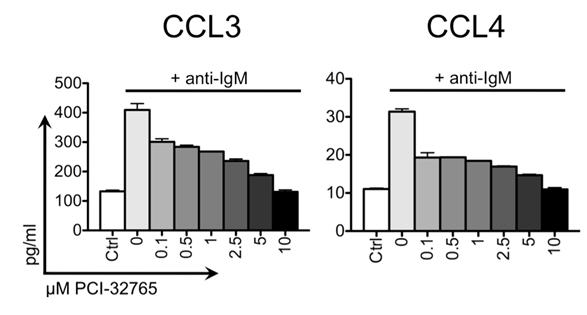 |
Figure: CCL3 and CCL4 concentrations in cell culture supernatants of the MCL cell line JVM-13 after 24 hour stimulation with anti-IgM and abrogation of this stimulation after pre-treatment with PCI-32765.
|